How nanocarriers deliver drugs inside cells (—from mystery to design)
Most of today’s most exciting drugs must work inside cells, yet many never make it past the cell’s own membrane-bound “security system”. My group uses advanced imaging and chemical biological reporters, to track how nano-sized drug carriers navigate the crowded cellular maze and to design carriers that do it better.
Text and photos Shiqi Wang
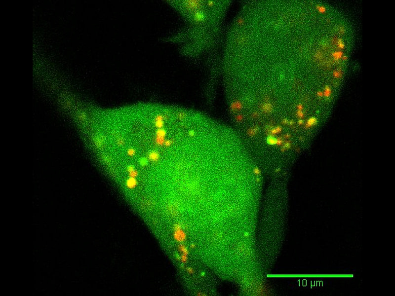
The fluorescent microscopy image of a cell, with endosomes labelled in red, and model drugs labelled in green. The uniform green in the cell means the drugs have escaped from endosomes.
I have always been fascinated by how cells interact with their surrounding environment. These tiny living creatures are the building blocks of our body. Every day, tens of trillions of cells work together, coordinate and create life. To me, this is a wonder.
From microscopes to unanswered questions
So when I pursued my PhD, I chose to study tiny nano-sized polymer particles, which can deliver fragile biological drugs into living cells. I spent many hours in front of microscopes, watching nanoparticles entering cells and disappearing eventually, wondering what really happened to them once they entered that crowded, complex world.
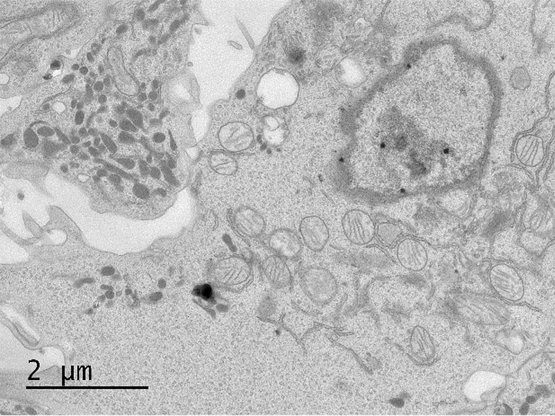
A transmission electron microscopy image of a cell, with bubble-like endosomes, striped mitochondria, and in the center a nucleus.
Well, I’m not the only person having those questions in mind. When cells swallow nanoparticles, they trap them in bubble-like compartments called endosomes and lysosomes—the cell’s sorting and recycling center. These compartments are designed to break down and recycle material, not to release delicate therapeutic cargo.
For a gene therapy or RNA drug to work, it must escape from these compartments into the cytosol, where the cell’s machinery can actually use it. This is called ”endosomal escape,” and it’s one of the biggest unsolved problems in nanomedicine.
Understanding endosomal escape is not an easy task.
Even the COVID-19 mRNA vaccines, which saved millions of lives, have shockingly low endosomal escape rates—current estimates suggest only a small percentage of the nanoparticles successfully release their cargo into the cytosol. They work because the dose is high and mRNA is potent, but they could work even better if we understood how endosomal escape happens and could improve the nanocarrier design accordingly.
Lighting up nanoparticles inside cells
Understanding endosomal escape is not an easy task. This is probably why we still don’t fully know how nanoparticles deliver drugs inside cells. There is an outline, but many details of the picture are missing.
I have loved solving puzzles since childhood, so I think, this is an interesting topic for my scientific career. To reveal those missing details, we need something beyond traditional microscopy to chase the nanoparticles inside cells, and to even get numbers showing how much drug truly arrives inside the cells where it can function.
A few years ago, I started thinking: what if we could design a reporter that only emits light when a drug molecule successfully reaches the cytosol?
A few years ago, I started thinking: what if we could design a reporter that only emits light when a drug molecule successfully reaches the cytosol? There are a few such biological reporters that exist but they are mainly based on split proteins. One piece is on the drug, and the other is expressed in the cytosol. Once the drug reaches cytosol, two complementary protein pieces form an active protein, and generate light signals.
I was thinking, probably we can do the same with smaller chemical reporters.
That’s when the idea of BioLure came up, splitting a light-emitting chemical into two pieces, and regenerating it through a so-called bioorthogonal reaction once the drug successfully reaches cytosol. This reaction can happen in live cells, without disturbing normal cell functions. We tested this idea on a cancer-killing protein delivery case, and we could directly link the amount delivered to how many tumour cells died.
This idea was first supported by the Research Council of Finland as a postdoctoral grant, and later I received the European Research Council Starting grant to further develop it as an assay, to measure endosomal escape.
The goal is ambitious: we want to create a platform where formulation scientists can test dozens of nanoparticle designs side by side and get numbers on which designs truly help drugs reach their destination, not just enter cells.
Chemistry, biology and Finland: my journey
When I started my PhD, non-viral gene therapy seemed like a distant dream. Then, during a global pandemic, I—and billions of others—received an mRNA vaccine delivered by lipid nanoparticles. The speed of that success was extraordinary, but it also revealed how much we still don’t understand.
Looking back, my scientific path has taken many turns since those early days at the microscope, yet they seem to converge on the interfaces of chemistry and biology. Living and working in Finland has also quietly shaped how I think about science. There is a certain calmness and trust in the research environment here that makes it easier to focus on long-term questions, even when the answers are not immediately clear. I have come to appreciate the openness of discussions across fields and the willingness to explore ideas that sit between disciplines. In many ways, that mirrors the kind of science I enjoy most—curious, collaborative, and not confined to a single boundary.
Introduction of the writer
Shiqi Wang is an Assistant Professor at the Faculty of Pharmacy, and Helsinki Institute of Life Science (HiLIFE), University of Helsinki. With a BSc and MSc in chemistry from China, a PhD in chemical engineering from the UK, and postdoctoral training at the University of Helsinki Faculty of Pharmacy, now Shiqi is leading a multidisciplinary research group, focusing on the fundamental understanding of biotherapeutics delivery into cells. She enjoys hiking, cooking, and exploring restaurants.